Test type
PGT-A, PGT-M or PGT-SR is selected according to the clinical question.

Preimplantation Genetic Testing (PGT) is a group of methods used during IVF to examine a limited sample of cells from an embryo for specific chromosomal or genetic risks.

PGT-A, PGT-M or PGT-SR is selected according to the clinical question.
An IVF/ICSI cycle is required to create embryos and perform a biopsy.
Blastocyst biopsy, sample identity verification, genetic analysis and embryo freezing may be used.
After the report and counselling, an embryo-transfer plan is prepared for embryos considered suitable for transfer.
PGT is an umbrella term for tests designed to answer a specific genetic question before embryo transfer.
A small number of cells taken from the embryo are analysed in a genetics laboratory. The scope depends on the selected type of PGT; the result is not a guarantee regarding the embryo’s entire genetic make-up.
PGT is not a blood test independent of IVF. Oocyte collection, fertilisation, blastocyst culture, biopsy and usually embryo freezing are required.
PGT-A screens numerical chromosome status. PGT-M is developed specifically for a known genetic change in a family. PGT-SR is designed to assess whether structural rearrangements in a parent, such as a balanced translocation or inversion, may result in unbalanced chromosome content in an embryo.
The result reflects the biopsied cell sample, the platform used and the laboratory’s reporting rules. Mosaicism, no-result findings and the possibility of misclassification should be discussed during counselling.
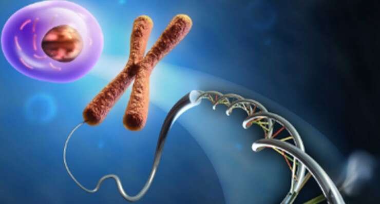
Although the tests may use the same biopsy sample, they answer different genetic questions.
Screens for missing or extra chromosome copies in an embryo.
PGT-M focuses on a single-gene condition, whereas PGT-SR focuses on unbalanced structural chromosome changes.
In some cases, PGT-A may be planned from the same biopsy sample alongside PGT-M or PGT-SR. The potential benefit, possibility of reducing the number of embryos available, cost and additional complexity of interpretation should be assessed individually.
The decision to use PGT is not made automatically based only on age or previous treatment failure.
A confirmed pathogenic genetic variant in the couple or family.
A balanced translocation, Robertsonian translocation or inversion identified in one parent.
Discussion of the potential benefits and limitations of PGT-A in selected patient groups.
Assessment of recurrence risk in families with a confirmed genetic diagnosis.
Targeted testing may be planned when clinical and genetic findings are appropriate.
The number of embryos expected to be frozen, age and future pregnancy goals are considered together.
No. In particular, routine PGT-A has not been shown to increase the chance of live birth for all IVF patients. The clinical question, potential benefit and possibility of reducing the number of embryos available should be defined clearly.
The genetic file and IVF plan should be completed as far as possible before ovarian stimulation begins.
The inheritance pattern, recurrence risk, testing options and alternatives are explained.
The laboratory checks whether the variant, karyotype or family diagnosis is suitable for test development or analysis.
DNA samples from relatives may be required for PGT-M test development.
Age, ovarian reserve, sperm status and expected blastocyst number are assessed.
Management of categories such as unaffected, carrier, affected, mosaic and no-result is discussed in advance.
Screening or diagnostic options that may be offered if pregnancy occurs are explained.
The process requires coordination of genetic preparation with IVF and embryology stages.
Family history, diagnostic reports and reproductive goals are reviewed.
PGT-A, PGT-M, PGT-SR or an appropriate combination is selected.
When required, a case-specific PGT-M assay is designed.
Oocytes are collected and fertilised using the appropriate method.
Embryos are monitored until they reach a developmental stage suitable for biopsy.
A limited number of trophectoderm cells are removed while maintaining sample identity and traceability.
The sample is analysed in the genetics laboratory using the selected method; embryos are usually frozen while awaiting results.
Results are interpreted with counselling and a frozen embryo-transfer plan is prepared for an appropriate embryo.
The timeline varies according to the test type and whether case-specific preparation is required.
Verification of existing reports can take days or weeks.
Depending on the case and family samples, this may take several weeks or longer.
Usually takes approximately 10–14 days before oocyte collection.
Usually performed on days 5–7 after fertilisation.
Varies according to the laboratory and test type.
A separate frozen embryo-transfer cycle may be planned after the result is available.
Some initial consultations and investigations can be organised remotely. Oocyte collection generally requires a plan of approximately 10–14 days; transfer may take place during a later, shorter visit.
Technical completion of the test is not the same as achieving pregnancy or live birth.
Affect the number of blastocysts reaching biopsy and the number of embryos potentially suitable after testing.
The chance that each embryo is affected or a carrier varies according to the inheritance pattern.
Not all fertilised oocytes reach the blastocyst or biopsy stage.
Insufficient DNA or technical issues can lead to a no-result report.
Mosaicism, segmental findings and laboratory thresholds can affect reporting.
An embryo considered genetically suitable for transfer still does not guarantee implantation or live birth.
Fertilisation, blastocyst development, test result, transfer, clinical pregnancy and live birth are separate stages. PGT provides information only about the selected genetic question.
Cost varies according to the IVF cycle, biopsy, test type and laboratory preparation required.
Ovarian stimulation, monitoring and oocyte collection.
Fertilisation and laboratory monitoring through the blastocyst stage.
Biopsy procedure, sample tubing and chain-of-identity documentation.
Scope of PGT-A, PGT-M or PGT-SR analysis and the number of embryos tested.
Case-specific preparation and family samples for PGT-M.
Embryo storage and the subsequent frozen embryo-transfer cycle.
Send us your genetic reports and previous IVF results so we can explain which test may be appropriate and itemise the likely costs.
Accurate identification of the biopsy sample from embryo to genetics laboratory is a fundamental component of reliable testing.
Patient, gamete, embryo, biopsy tube and report are verified within the same chain of identity.
A limited number of trophectoderm cells are removed by a trained embryologist.
Cells are transferred into a tube using a controlled workflow designed to minimise DNA loss and sample mix-up.
Following biopsy, embryos are stored using rapid freezing while results are pending.
Embryo codes and result categories are entered into the clinical record using double verification.
Equipment, operator, date, sample location and report amendments are documented.

IVF treatment, embryo biopsy and clinical interpretation of the genetic report require collaboration between different specialties.

Coordinates ovarian stimulation, oocyte collection and transfer planning with the genetic objective.
View profile →
Coordinates genetic testing, consent, multidisciplinary workflow and scientific quality processes.
View profile →
Manages embryo culture, biopsy, vitrification, identity verification and matching of the report to the correct embryo.
View profile →Clinical interpretation of genetic risk and test results should involve an appropriately qualified medical geneticist or genetic counsellor. The IVF and embryology team manages integration of testing into reproductive treatment.
PGT may aim to reduce a specific genetic risk, but it has biological and technical limitations.
Not all embryos reach the blastocyst stage or are considered suitable for transfer after testing.
Insufficient DNA or technical reasons may lead to consideration of re-biopsy.
Biopsied cells may not always represent the entire embryo.
Although uncommon, false-positive or false-negative results are possible.
Although risk is low in experienced laboratories, zero risk to the embryo cannot be promised.
Genetic conditions not included in the test, de novo changes and pregnancy complications are not excluded.
Key answers about PGT types, the process, results and follow-up during pregnancy.
A group of tests used to assess specific chromosomal or genetic risks before embryo transfer.
No. PGT-A assesses chromosome number, PGT-M a specific single-gene condition and PGT-SR structural chromosome imbalance.
Yes. An IVF or ICSI cycle is required to create embryos and perform a biopsy.
In current practice, it is most commonly performed at the blastocyst stage, usually on days 5–7.
The risk is low in experienced laboratories, but zero risk from biopsy and freezing cannot be guaranteed.
No. It evaluates only the genetic question within the scope of the selected test.
Rare incorrect results, mosaicism and no-result analyses are possible.
Because PGT does not exclude every fetal genetic condition, screening and, when appropriate, diagnostic options such as CVS or amniocentesis should be discussed during pregnancy.
They are usually vitrified after biopsy and transfer is planned after results are available.
The purpose varies according to the test type and patient group. In particular, PGT-A has not been shown to increase live birth for every patient.
There is no fixed number. Age, genetic risk and embryo development influence the chance of finding an embryo suitable for transfer.
The IVF part usually takes approximately 10–14 days; genetic preparation and reporting times vary separately according to the test type.
IVF, medication, biopsy, testing, freezing, storage and transfer may be charged as separate items.
It refers to a biopsy result suggesting a mixture of cells with normal and abnormal chromosome copy-number findings; interpretation and transfer policy require specialist counselling.
Message us on WhatsApp for information about treatments, the process and planning your visit to Cyprus.