
EmbryoScope+ Embryo Monitoring
Embryo development changes hour by hour. EmbryoScope+ combines incubation with time-lapse imaging so developmental images can be recorded at defined intervals while embryos remain within a controlled culture environment.
- Less routine handlingEmbryos can be observed through recorded images without routine removal for every assessment.
- Morphokinetic dataDivision timing and developmental patterns can be reviewed over time.
- Retrospective reviewRecorded sequences allow unusual developmental events to be re-examined.
Time-lapse monitoring is a laboratory observation tool. It does not guarantee implantation, pregnancy or live birth and does not replace the embryologist’s assessment.
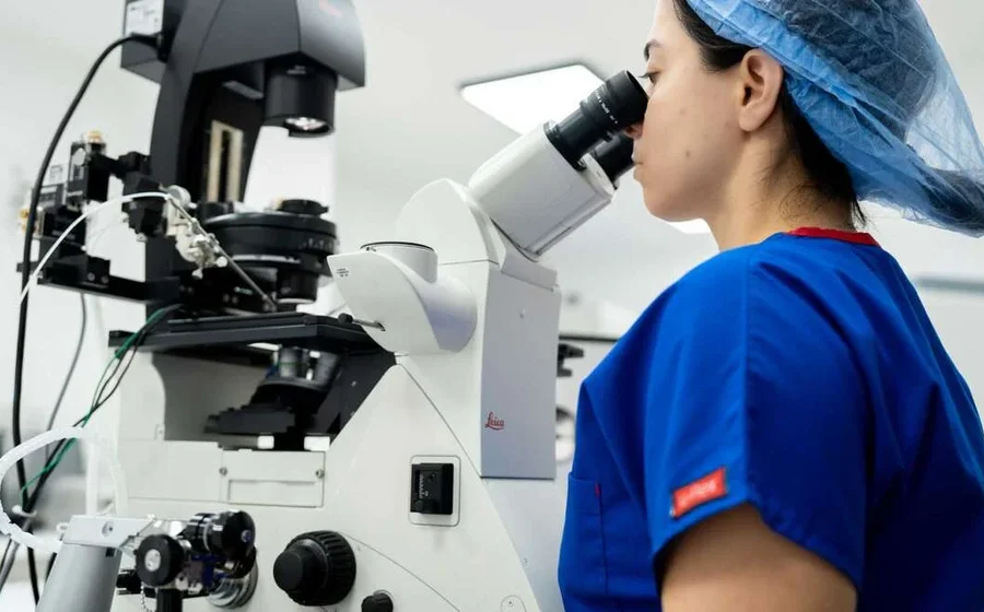 Time-lapse · Developmental recording
We want important moments in embryo development to be available for review, not missed between routine checks.
Embryo development is dynamic. The timing and sequence of cell divisions and progression towards the blastocyst stage may provide information that is not visible in a single snapshot.
We use time-lapse technology as a culture and observation system that can provide richer developmental information while keeping clinical interpretation with the embryology team.
Reduced external handling
Routine visual checks can be performed from recorded images rather than repeatedly moving embryos for observation.
Morphokinetic information
The timing and order of key developmental events can be documented.
Retrospective review
Recorded sequences can be reviewed when an unusual developmental pattern is seen.
Structured data
Time-lapse records may support standardised assessment and, in validated systems, AI-assisted analysis.
What is EmbryoScope+ time-lapse embryo monitoring?
EmbryoScope+ is an incubator with an integrated imaging system. It captures embryo images at programmed intervals and creates a chronological developmental record while embryos remain in culture.
The system allows embryologists to review morphology together with timing information. It does not make the transfer decision by itself; the record is interpreted alongside embryo quality, developmental day, clinical history and any relevant genetic information.
Important distinction: more images do not automatically mean a better clinical outcome. The value of time-lapse lies in observation, documentation and decision support within a validated laboratory workflow.
Time-lapse monitoring adds the dimension of time to conventional embryo morphology.
An embryo may look similar at two observation points while having followed different developmental paths between them. A continuous record can help the embryology team recognise timing patterns and unusual cleavage events.
Fertilisation
Normal fertilisation is confirmed and the developmental record begins.
Culture
Embryos remain in a controlled incubator environment while images are captured.
Timeline
Cell divisions and progression are organised into a developmental sequence.
Interpretation
The embryologist reviews morphology, timing and the overall clinical plan together.
How does EmbryoScope+ monitoring work step by step?
The exact imaging interval and culture protocol depend on the laboratory’s validated procedures. The central principle is to create a developmental timeline while minimising unnecessary disturbance.
Fertilisation is assessed
Normal fertilisation is documented and embryos suitable for culture are identified.
Embryos are placed in the system
Embryos are cultured under controlled temperature and gas conditions.
Images are captured automatically
The integrated camera records images at defined intervals without routine removal for imaging.
Developmental events are annotated
Division timing and relevant morphokinetic events can be reviewed.
The embryologist interprets the record
Time-lapse information is considered together with standard morphology and clinical information.
Transfer or cryopreservation is decided
The final plan is made according to embryo development and the personalised treatment strategy.
What can be reviewed in a time-lapse embryo record?
The exact variables used vary by laboratory and software. Time-lapse data should not be interpreted as a universal scoring system.
Developmental timing is one information layer. Morphology, genetic results when available and the patient’s clinical context remain important.
Cleavage timing
The timing of transitions between early cell stages can be documented.
Cleavage pattern
Direct, reverse or otherwise unusual division patterns may be identified.
Fragmentation and symmetry
Visible structural changes can be reviewed across multiple time points.
Blastocyst formation
The timing and progression of blastulation can be documented.
Developmental arrest
Periods of slowed or arrested development can be reviewed retrospectively.
Complete image sequence
The embryologist can return to earlier time points when assessing the developmental story.
Routine observation and time-lapse monitoring
Time-lapse technology can strengthen developmental observation, but evidence for a universal improvement in live-birth outcomes remains context dependent.
Professional guidance recognises time-lapse systems as tools for embryo culture, imaging and developmental assessment. Studies have examined morphokinetic markers and selection algorithms, but findings are not interchangeable across every system and population.
The most defensible use is therefore structured observation and decision support within a validated laboratory process, rather than presenting the technology as a stand-alone success intervention.
A time-lapse record can provide more developmental context, but embryo prioritisation should still be integrated with morphology, clinical factors and genetic information when available.
Why do we state the limits clearly?
More data is not a guarantee. A richer image record does not ensure implantation or live birth.
Algorithms are system-specific. A score developed for one platform or population should not automatically be generalised.
Genetic status is separate. Time-lapse appearance does not establish chromosomal normality.
Culture quality still matters. Imaging cannot replace good incubator performance, media control and laboratory practice.
When may time-lapse embryo monitoring be useful?
The technology can be used across many IVF cycles when the laboratory’s culture strategy supports it. Its added value depends on the clinical question and the number and development of available embryos.
Blastocyst culture
A longer developmental record can be reviewed as embryos progress towards the blastocyst stage.
Several embryos
Timing and morphology can provide additional information when prioritising among available embryos.
Previous cycle review
Recorded development may help the team review unusual cleavage or arrest patterns.
AI-assisted assessment
Time-lapse sequences can supply data to validated image-analysis systems where available.
PGT cycles
Developmental records can be interpreted alongside biopsy and genetic results.
Laboratory quality review
Recorded sequences can support retrospective review and training.
What does this technology not do?
Clear limits help prevent unrealistic expectations and keep laboratory information in the correct clinical context.
It does not guarantee that an embryo will implant or result in a live birth.
It does not provide a genetic diagnosis and does not replace PGT when clinically indicated.
It does not make the final embryo-transfer decision independently.
A time-lapse score should not automatically be used to discard an embryo.
Its value depends on validated culture, imaging, software and embryology procedures.
At Ventus IVF, the timeline supports the embryologist; it does not replace the embryologist.
We interpret time-lapse records together with morphology, developmental day, any available genetic result and the patient’s treatment plan.
When a developmental pattern is unusual or a score conflicts with the wider picture, the specialist team reviews the record rather than relying on a single automated output.
Ask about your situationCommon questions about EmbryoScope+ Embryo Monitoring
These answers provide general laboratory information and do not replace personalised medical advice.
Ask about your situationDoes EmbryoScope+ watch embryos continuously?
The system captures images at programmed intervals and creates a time-lapse sequence that can be reviewed throughout culture.
Are embryos removed from the incubator for every check?
The integrated imaging system is designed to reduce the need for routine removal solely for visual observation.
Does EmbryoScope+ guarantee a better embryo?
No. It records development; it does not change an embryo’s underlying biological potential.
Can time-lapse show whether an embryo is genetically normal?
No. Morphology and developmental timing are not a genetic diagnosis.
Can AI use EmbryoScope+ images?
Some validated systems can analyse time-lapse images, but the available software and intended use depend on the laboratory platform.
Can a lower time-lapse score still result in pregnancy?
Yes, a score is not an absolute outcome. Final prioritisation should consider the complete clinical and embryology picture.
Explore related technologies
Different laboratory technologies support different parts of assessment, safety, culture and cryopreservation.
Scientific sources and further reading
- ESHRE recommendations for good practice for the use of time-lapse technology. Source ↗
- Vitrolife EmbryoScope+ time-lapse system product information. Source ↗
- Deep learning versus manual morphology-based embryo selection in IVF. PubMed PMID: 39122964. Source ↗
- ASRM committee opinion on artificial intelligence in the IVF laboratory. Source ↗
Ask whether time-lapse embryo monitoring may add useful information to your treatment plan.
Our team can explain how embryos are cultured and monitored, how developmental records are interpreted and where the limitations of time-lapse technology remain.